






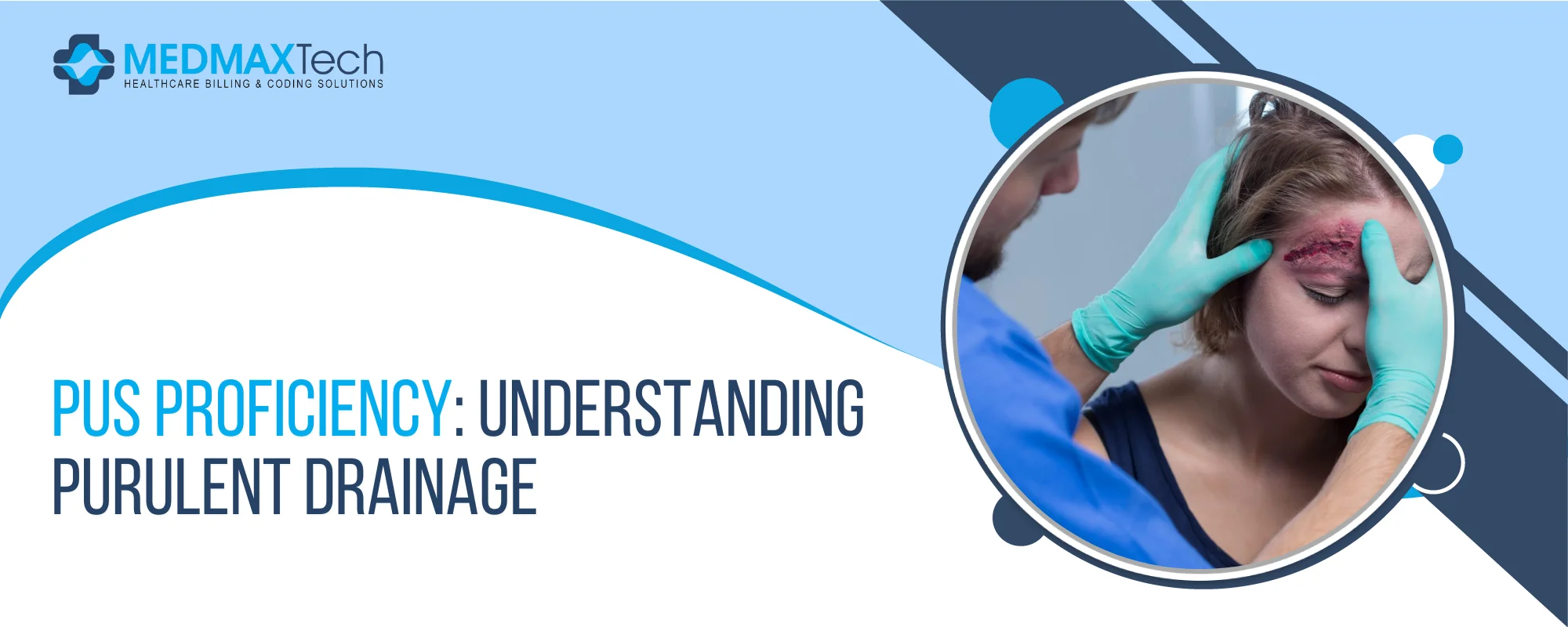
Pus Proficiency: Understanding Purulent Drainage
Wound drainage management is a very important part of wound care. The presence of purulent drainage helps show whether an infection is present and how the wound is healing. Non-healing wounds affect millions of people in the United States, especially many adults over the age of 65. Understanding the characteristics, meaning, and proper management of purulent drainage is essential for achieving the best possible wound healing.
Purulent Drainage: The Basics
Characteristics
Purulent drainage, also known as pus, is a thick and cloudy fluid produced by the body during an inflammatory response to infection. It commonly contains dead white blood cells, bacteria, tissue debris, and proteins that build up while the body fights infection.
Pus may appear cream-colored, yellow-green, brown, or even pink-tinged if blood or other pathogens are present. In most cases, there is some level of odor, which can range from mild to very strong.
Mechanism
Pus forms as part of the body’s immune response when pathogens invade. When bacteria break through the skin’s protective barrier, neutrophils (specialized white blood cells) are activated. They perform phagocytosis, which means they consume and destroy the invading pathogens, and then they die. The buildup of these dead neutrophils along with other debris creates the appearance of pus.
The buildup of dead neutrophils, bacteria, and tissue debris creates pus. This sequence of events is known as the inflammatory cascade:
- Pathogen enters the body and triggers an immune response
- Neutrophils move to the wound site
- Phagocytosis occurs
- Dead neutrophils and debris collect
- Purulent drainage forms
Clinical Significance
The presence of pus usually signals an infection and often requires medical care. However, not every case of purulent drainage means a severe infection. The wound’s condition, the amount of drainage, and related symptoms must all be evaluated.
Signs that suggest medical treatment is needed include:
- Volume changes: An increase in the amount of pus may show that the infection is getting worse.
- Color transitions: Normal wound drainage is pale or slightly tinted. Purulent drainage is thick and milky. Green, yellow, or brown pus may signal infection progression.
- Consistency: Normal wound drainage is thin, while purulent drainage is much thicker.
- Associated wound characteristics: A normally healing wound should have little to no odor. A foul smell may indicate infection. The surrounding skin may feel warm or show red streaks, which can suggest cellulitis.
- Systemic symptoms: Tenderness, fever, and a high white blood cell count may occur when infection is present.
Assessment and Management of Purulent Drainage
Visual Examination
Basic wound assessment principles should always be followed:
- Identify the wound type and cause
- Assess the patient’s overall health to understand healing potential. Elderly patients, diabetics, and immunocompromised individuals often heal more slowly
- Document wound size, depth, and location
- Check for infection. Drainage alone does not always mean infection, but the appearance and type must be documented
Common types of wound drainage include:
- Sanguineous: Seen shortly after injury and contains fresh blood. It appears bright red or pink and should decrease
- quicklSanguineous: This type appears in the first few hours after an injury. It mainly contains fresh blood and looks pink or bright red. In a full-thickness or partial-thickness wound, it can continue for a few days after the injury, but it should reduce quickly.
- Sanguineous: This type appears in the first few hours after an injury. It mainly contains fresh blood and looks pink or bright red. In a full-thickness or partial-thickness wound, it can continue for a few days after the injury, but it should reduce quickly.
- Serosanguineous: This is a mix of serous and sanguineous drainage and is the most common type of discharge. It usually looks pale and thin with a slight reddish color. It often indicates that an infection is clearing.
- Serous purulent drainage: This thin and light-colored drainage mostly contains white blood cells. It appears during the inflammatory wound healing stage, usually within the first 48–72 hours after an injury.
- Make sure the wound is moving properly through the wound healing stages.
- Hemostasis – A fibrin clot forms to stop acute bleeding.
- Inflammation – Neutrophils and macrophages become active to fight invading microbes, releasing cytokines and other inflammatory agents.
- Proliferation – Cell growth and activity create new skin and blood vessels.
- Remodeling – The skin heals, and the extracellular matrix reshapes and strengthens itself.
- Reassess the wound on a regular basis. All wounds, especially infected wounds, should be checked frequently by the care team. Any clear or unusual changes should be documented, and the appropriate healthcare team member should be informed right away.
Measurement and Diagnosis
Culturing Wounds
Proper diagnosis and management of infections are very important for wound healing. All open wounds contain bacteria, and healing can still happen even when bacteria are present. Many types of aerobic and anaerobic bacteria can contaminate wounds. However, the type of bacterial species is more important than the number of bacteria.
There are two common techniques used to swab a wound for culture: the Levine technique and the Z technique. In the Levine technique, the sample is taken from a smaller, specific area of the wound. The swab is rotated with enough pressure to draw out fluid from the tissue. Recent studies show that the Levine technique is preferred when collecting bacterial cultures.
In the Z technique, the swab is turned between the fingers and moved in a zigzag pattern across the wound surface without touching the wound edges.
Other Diagnostic Methods
Although a culture swab is still the main method used to diagnose bacterial colonization in a wound, other tests may be required depending on the patient, the wound location, and how severe the wound is.
- Wound biopsies can be very helpful in diagnosing invasive burn wounds. If bacteria are found in the deep dermis during a biopsy, it is linked to a higher risk of sepsis in a burn patient. A bone biopsy is considered the gold standard for diagnosing osteomyelitis.
- Laboratory studies can also support the management of chronic wounds, but they are often not needed because routine blood work is usually not required. In some cases, tests such as serum prealbumin can be used to assess nutritional status. Erythrocyte sedimentation rate (ESR) and C-reactive protein (CRP) may help confirm a diagnosis of osteomyelitis or monitor how well the treatment is working.
- Imaging tests like ultrasound, computed tomography (CT), or magnetic resonance imaging (MRI) can be useful in certain situations to identify fluid collections, necrotic tissue, or areas of inflammation.
Assessment and Documentation
Since wounds are usually managed by a multidisciplinary team, keeping consistent records is very important to share information with all team members. There are many ways to document wounds, but a few standardized tools can be especially useful in practice. These tools help not only with the initial diagnosis, as mentioned earlier, but also during follow-up checks to track the wound’s progress over time.
Bates Jensen Wound Assessment Tool
This method evaluates 15 aspects of a wound, including its size, depth, edges, undermining, type and amount of necrotic tissue, type and amount of exudate, surrounding skin color, peripheral edema and induration, granulation tissue, and epithelialization. Out of these, 13 items are scored, while the location and shape of the wound are not scored. Each scored item is rated on a scale from 1 to 5, with 5 representing the most severe condition. The scores are added together for a maximum total of 65, and this total can be used to track the wound’s progress over time.
TIMERS
This observation-based tool focuses on:
- Tissue management
- Infection/inflammatio
- Moisture
- Epithelial edge
- Repair/regeneration
- Social factors
REEDA
Although mainly used for perineal wounds, REEDA evaluates redness, swelling, bruising, discharge, and wound closure and may be applied to other wounds.
AI-powered wound imaging software allows providers to compare wound images with large databases, improving diagnosis and treatment tracking.
Treatment for Purulent Drainage
Treatment of purulent wounds should begin quickly to prevent complications. The approach depends on the wound’s size, location, healing stage, and the patient’s overall health. The main goals are to stop further damage and keep the wound clean to prevent more infection.
The choice of dressing and cleaning method will depend on the type of infection. The patient’s overall risk must also be assessed. For example, wounds caused by venous insufficiency may need elevation and compression to manage drainage. Deeper or chronic wounds may benefit from negative pressure wound therapy.
Doctors may prescribe antibiotics, and if an abscess forms, it might need to be drained or surgically removed to help the wound heal properly.
Complications
Untreated wound infections can spread and cause serious problems:
- Cellulitis: affects the deeper layers of the skin. It usually shows as warmth and red streaks near the wound. Other symptoms may include fever, lightheadedness, nausea, and vomiting.
- Osteomyelitis: is a bacterial infection of the bone. It causes pain, swelling, and redness around the affected area. Patients may also have fever and feel very tired.
- Sepsis: happens when bacteria enter the bloodstream, causing a serious, widespread infection. This is a medical emergency and needs immediate hospitalization.
- Necrotizing soft tissue wounds: are rare but extremely dangerous. They are classified based on the soft tissue layer affected and include necrotizing cellulitis, necrotizing fasciitis, and necrotizing myositis. These conditions require urgent medical attention.
The key signs of this disease, such as blisters (bullae), a crackling feeling under the skin (crepitus), gas or air seen on X-ray, low blood pressure (hypotension), and dead skin (skin necrosis), are rarely visible when the patient first appears. Therefore, the diagnosis is mostly based on clinical evaluation by the healthcare provider.
- C-reactive protein (CRP)
- White blood cell count (WBC)
- Hemoglobin
- Sodium
- Creatinine
- Glucose
Even with these tools, if necrotizing fasciitis is strongly suspected, surgical removal of infected tissue (debridement) should be done immediately without waiting for test results.
Multidisciplinary Approach
Caring for infected wounds needs a team approach. This often includes not just the wound care specialists, but also surgeons, infectious disease experts, and physical therapists. An important but sometimes overlooked team member is the patient themselves, as actively participating in their own care can bring many benefits.
Improved Self-Management
Teaching patients about their wounds helps them take an active role in their care. When they understand the wound and the importance of keeping it clean, healing improves. Patients who are well-informed are more likely to follow the care instructions properly.
Prevention of Complications
Taking proper care of wounds can lower the risk of problems like infections and slow healing. Teaching patients to recognize signs of infection or other issues helps them get treatment earlier.
Healthy Lifestyle Choices
This is a great time to educate patients on how their lifestyle affects healing. Advising them on healthy eating, quitting smoking, and managing chronic conditions like diabetes can greatly improve treatment outcomes.
Superior Surgical Wound Care
For surgical wounds, the basics like keeping the incision clean and dry are important for almost all patients. Additionally, each patient will receive wound care tailored to their specific needs to support proper healing.
Improved Communication
When patients understand their care, they can communicate their progress more effectively with healthcare providers. Clear communication helps them follow treatment plans, take medications correctly, and attend follow-up appointments. This teamwork is key for a successful recovery.
Reduced Healthcare Costs
Better education leads to fewer complications, hospital stays, and treatments.
Managing Purulent Drainage: Today and in the Future
Most purulent drainage care still follows basic steps: assessment, culturing, proper cleaning, dressing, and treatment. A team-based approach remains essential.
New treatments such as bacteriophage therapy, growth factors, bioengineered tissues, and stem cell treatments show promise. Advanced dressings and technologies may further improve wound healing in the future.






Williams Clark
Williams Clark is a dual-certified healthcare professional with CPC and CHC credentials. With over a decade of experience in medical billing and compliance auditing and provides deep insights into payer policies, denial prevention, and accurate coding practices. His mission is to empower medical billers and healthcare providers with the knowledge to stay compliant and profitable.